
Gundersen HJ et al (1988) The new stereological tools: disector, fractionator, nucleator and point sampled intercepts and their use in pathological research and diagnosis. Gardella D, Hatton WJ, Rind HB, Rosen GD, von Bartheld CS (2003) Differential tissue shrinkage and compression in the z-axis: implications for optical disector counting in vibratome-, plastic- and cryosections. doi: 10.1016/j.reprotox.2005.04.006ĭorph-Petersen KA, Nyengaard JR, Gundersen HJ (2001) Tissue shrinkage and unbiased stereological estimation of particle number and size. doi: 10.2478/s1338-8ĭe Groot DM et al (2005) 2D and 3D assessment of neuropathology in rat brain after prenatal exposure to methylazoxymethanol, a model for developmental neurotoxicty. doi: 10.1186/s1298-8īrasnjevic I et al (2013) Region-specific neuron and synapse loss in the hippocampus of App/PS1 knock-in mice.

doi: 10.1002/jemt.20345īrandenberger C, Ochs M, Muhlfeld C (2015) Assessing particle and fiber toxicology in the respiratory system: the stereology toolbox.

J Microsc 196:69–73īaryshnikova LM, Von Bohlen Und Halbach O, Kaplan S, Von Bartheld CS (2006) Two distinct events, section compression and loss of particles (“lost caps”), contribute to z-axis distortion and bias in optical disector counting.

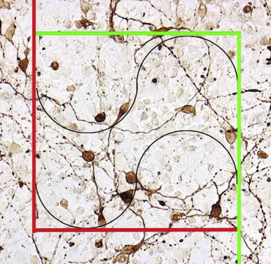
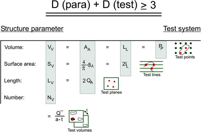
Furthermore, it is described how to perform immuno-histochemical staining of such thick cryo-sections, followed by providing a guidance for quantification of the ROI volume, the generation of unbiased virtual counting spaces, and steps to work with these counting spaces to obtain an unbiased estimate of total cell numbers.Īndersen BB, Gundersen HJ (1999) Pronounced loss of cell nuclei and anisotropic deformation of thick sections. Steps described in this protocol include transcardial perfusion of mice, postfixation, and cryoprotection of the region of interest (ROI), followed by the preparation of a systematically and randomly sampled series of thick sections through the entire ROI. Design-based stereology has become the method of choice for unbiased, reproducible total cell number quantification. The number of a specific cell type cannot be simply deduced from the number of its profiles found in thin tissue sections, as this parameter also depends on cell volume, tissue orientation as well as tissue atrophy. Valid quantification of organ volume and total cell numbers are crucial parameters for morphometric studies.


 0 kommentar(er)
